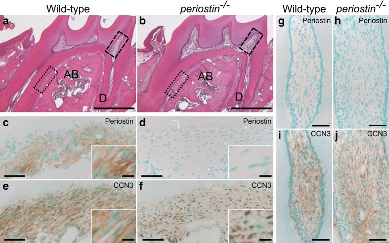
Fig. 5.
Decreased matricellular localization of CCN3 in the periostin -deficient periodontal ligament. Hematoxylin and eosin staining (a, b) and immunohistochemical analysis (c–j) of the mandibles of the mice. Paraffin sections of mandibles from 8-week-old wild-type (a, c, e, g, i) and periostin −/− (b, d, f, h, j) mice were stained with hematoxylin and eosin (a, b), anti-periostin (c, d, g, h), or anti-CCN3 (e, f, i, j) antibodies. High magnifications of dotted-line boxes and dashed-line boxes in “a” and “b” indicate the periodontal ligament ( c–f) and the dental pulp (g–j), respectively. Typical immunoreactivities in “c”-“f” are represented as high-magnification images in the insets (white-lined boxes). AB, alveolar bone; D, dentin. Scale, 500 μm (a, b); 50 μm (c–f); 20 μm (insets of c–d); 100 μm (g–j)

