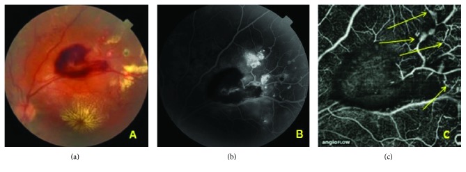
Figure 1.
Case 1: central and peripheral lesions. (a) Color fundus photograph of the left eye. The upper temporal quadrant shows a large hemorrhage; lipid deposits in the macula and superior to the hemorrhage. (b) Late-phase FA. Multiple vascular anomalies in the upper temporal quadrant of the midperiphery of the retina. The vascular abnormalities located more peripherally are visible only in FA. (c) OCTA image of vessels in the upper temporal region. Superficial plexus. Vascular anomalies, dilated vessels and loops (arrows). The retinal blood flow is partially obscured due to hemorrhage.

