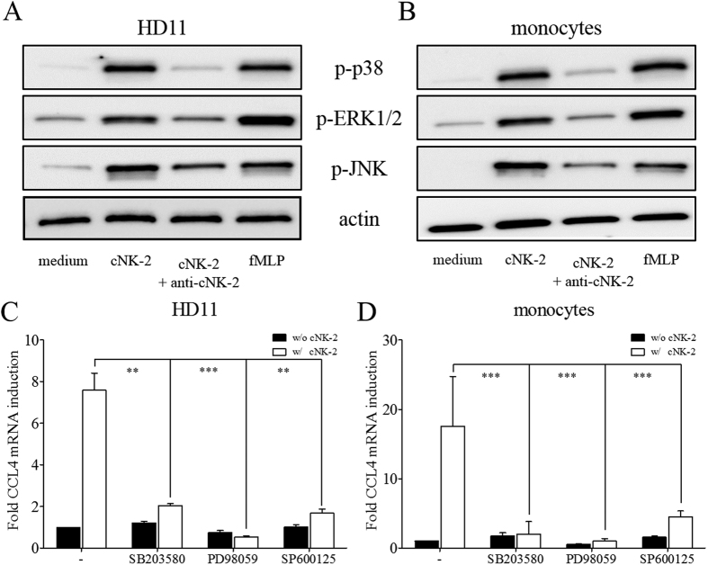
Figure 4. Activation of the MAPK pathway by cNK-2.
HD11 cells (A) and primary monocytes (B) were stimulated with 10 μg/ml cNK-2 or fMLP as a positive control for 30 min. The levels of phosphorylated p38 (p-p38), phosphorylated ERK1/2 (p-ERK1/2), phosphorylated JNK (p-JNK) and β-actin in the cellular lysates were determined by Western blot analysis. The effects of MAPK inhibitors (10 μM each) on p38 (SB203580), ERK1/2 (PD98059) and JNK (SP600125) in HD11 cells (C) and primary monocytes (D) were determined by real-time qPCR. p < 0.01 (**) and p < 0.001 (***) were considered statistically significant compared to the vehicle control.

