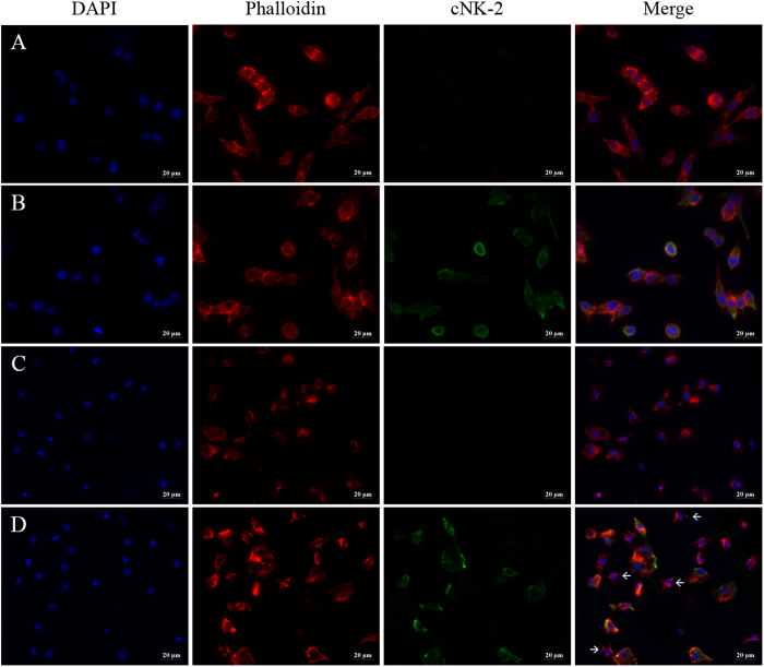
Figure 5. Cellular localization of cNK-2.
HD11 cells (A and B) and primary monocytes (C and D) were incubated with medium alone (A and C) or 10 μg/ml cNK-2 (B and D) for 30 min at 41 °C. Immunocytochemistry was performed with a rabbit polyclonal cNK-2 (green) antibody followed by an Alexa Fluor 488 goat anti-rabbit IgG secondary antibody. DAPI and Alexa Fluor 555 Phalloidin were used to stain nuclei (blue) and F-actin (red), respectively. The scale bar in all figures corresponds to 20 μm.

