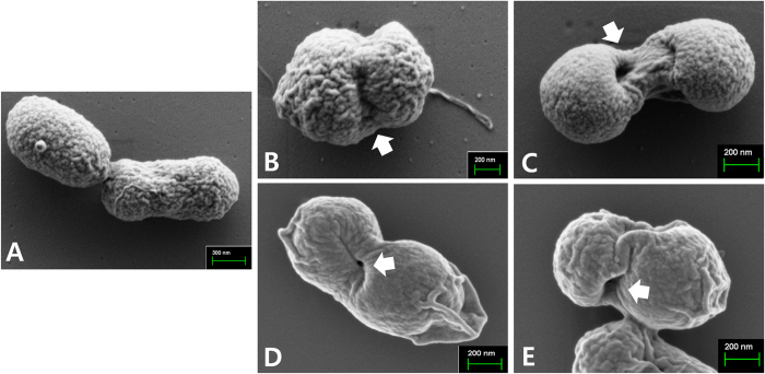
Figure 4. Characterization of JOL1954 S. Typhimurium ghost cells by scanning electron microscopy (SEM).
(A) Intact JOL1954 cells before the lysis. (B) JOL1954 ghost cells after 12 hr of the lysis. (C) JOL1954 ghost cells after 24 hr of the lysis (D) JOL1754 ghost cells after 12 hr of the lysis (E) JOL1754 ghost cells after 30 hr of the lysis. The arrows indicate the lysis transmembrane tunnels.

