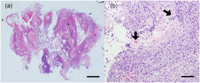
Figure 2.
Histopathology of the tissue easily removed by gentle manipulation. (a) At a low magnification degenerative changes, such as necrosis and vacuolar and hydropic degeneration, are seen, but there are areas of slight or no degeneration that allow observation of pathological findings. Severe papillomatous hyperplasia is seen in the epidermis, and the external root sheaths of hair follicles are preserved. Haematoxylin and eosin, bar = 500 μm. (b) At a higher magnification, the epidermis shows parakeratotic hyperkeratosis, with infiltrating cells that are mainly neutrophils and lymphocytes. Apoptotic keratinocytes are also observed (arrows). Haematoxylin and eosin, bar = 50 μm

