
Figure 3.
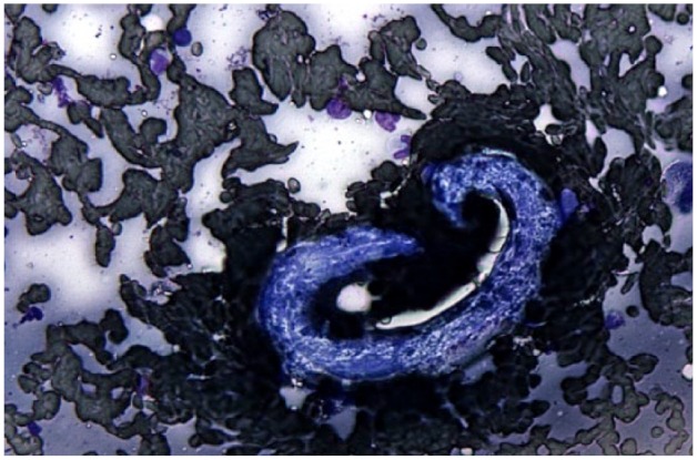
Cytologic sample obtained via pulmonary ultrasound-guided fine-needle aspirate containing Aelurostrongylus abstrusus larva. Just to the right of the center of the image is a large, curved, basophilic larva with a kinked tail (far right) and non-rhabditiform esophagus (left) extending approximately half of the length of the parasite, consistent with a stage 1 A abstrusus larva. The background consists of blood and proteinaceous debris interspersed with occasional non-degenerate neutrophils and alveolar macrophages. Modified Wright’s stain