
Figure 4.
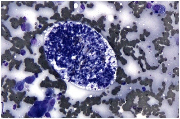
Cytologic sample obtained by way of ultrasound-guided pulmonary fine-needle aspiration showing A abstrusus ova. At the center of the image is a large, oval egg containing numerous basophilic-staining blastomeres surrounded by abundant non-degenerate neutrophils and alveolar macrophages (characterized by their abundant, vacuolated cytoplasm) on a background of blood and proteinaceous debris. Modified Wright’s stain