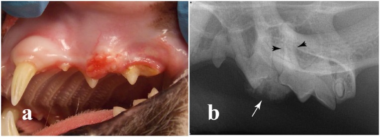
Figure 1.
(a) A 10-year-old domestic shorthair cat presented with an erythematous, firm ulcerated gingival mass on the buccal aspect of the left maxillary third and fourth premolar teeth (teeth 207 and 208). (b) The corresponding intraoral dental radiograph showed a well circumscribed, radiopaque mass (arrow) affecting the interproximal region between teeth 207 and 208; some tooth resorption and loss of the periodontal ligament space (arrowheads) are evident around the mesiobuccal root of tooth 208

