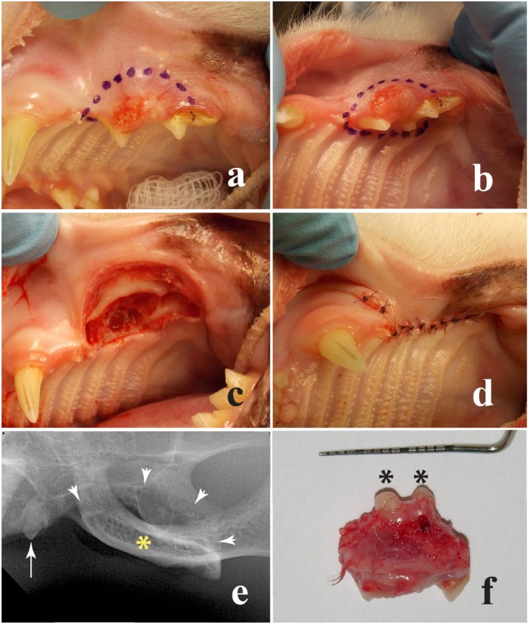
Figure 2.
(a) Buccal and (b) ventral views to the left maxillary area in a 10-year-old domestic shorthair cat. A surgical marker pen was used to outline the planned incisions. (c) Intraoperative view of the affected area after excision of the mass. (d) Closure buccal flap was sutured over the wound, using an absorbable monofilament in a simple interrupted pattern. (e) A postoperative intraoral radiograph was obtained, showing the affected area after rim excision (arrowheads), the left maxillary second premolar (arrow) and the zygomatic arch (asterisk). (f) The excised tissue was submitted for histopathological assessment. The asterisks show some intact roots of the third and fourth premolar teeth. Remaining root tips were extracted following excision of the mass. Because this was a clean-contaminated procedure, no surgical drapes were used. However, aseptic technique, including disposable synthetic or reusable cloth drapes may be used to minimize contamination6,7

