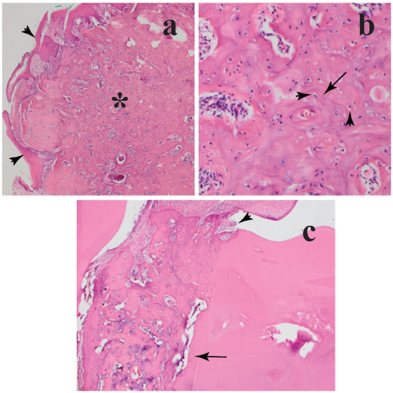
Figure 3.
(a) Photomicrograph of the submitted specimen showing proliferation of mineralized eosinophilic material (asterisk) that expands the gingiva (arrowheads) (×4 magnification, hematoxylin and eosin). (b) There are multiple irregularly placed lacunae (arrowheads) and reversal lines (arrow) (×20 magnification, hematoxylin and eosin). (c) The mass is focally adhered to a tooth root near the cementoenamel junction (arrowhead), contiguous with the normal cementum layer (arrow) (×4 magnification, hematoxylin and eosin)

