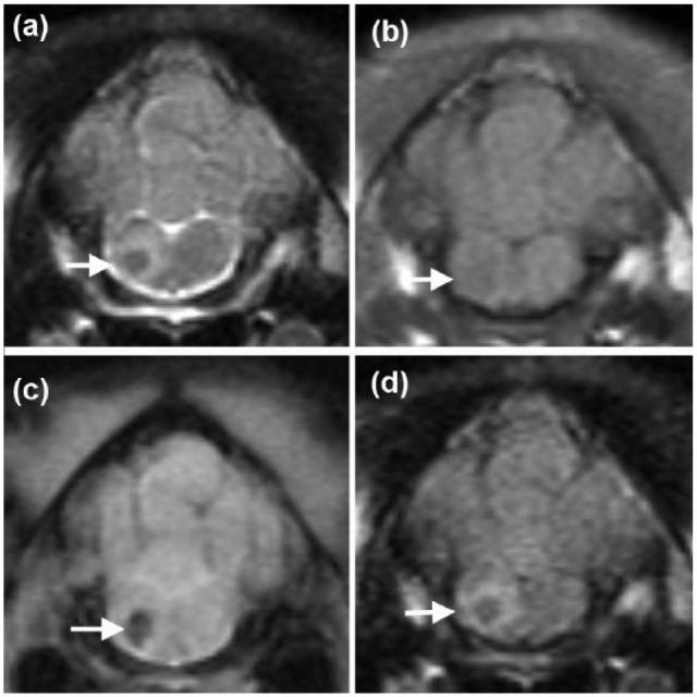
Figure 1.

Transverse magnetic resonance images of the head of a 6-month-old female domestic shorthair cat at the level of the myelencephalon, illustrating the well-defined lesion that is hypointense on (a) T2-weighted and iso- to hypo-intense on (b) T1-weighted images, with a signal void on (c) T2*-weighted images and a perilesional rim hyperintensity on (a) T2-weighted and (d) fluid attenuated inversion recovery images (arrows)
