
Figure 2.
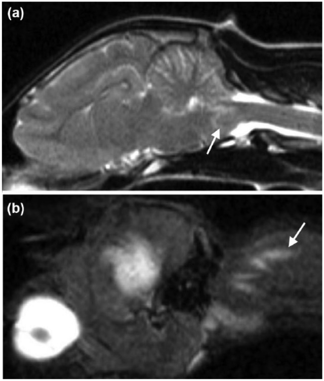
Sagittal magnetic resonance images of the head and neck of a 6-month-old female domestic shorthair cat illustrating the hypointense lesion surrounded by a hyperintense perilesional rim on T2-weighted images (a, white arrow). On the parasagittal view, a caudodorsal to cranioventral linear hyperintense lesion in the muscles of the dorsolateral aspect of the neck and in the direction of the cisterna cerebellomedullaris on short tau inversion recover images was observed (b, white narrow)