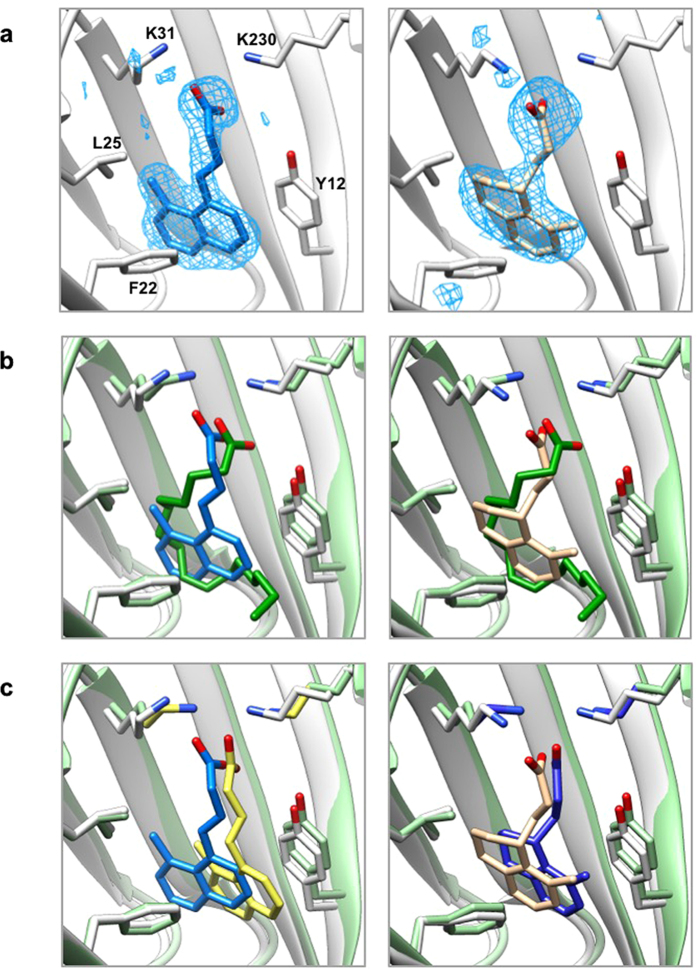
Figure 6. X-ray crystal structures of ToxT-inhibitor complexes reveal compounds bind in the fatty acid-binding pocket.
(a) Simulated annealing Fo – Fc omit maps of ToxT bound to compounds 3b (left) and 5a (right) contoured at 2.5 σ. (b) Overlay of crystal structures of ToxT bound to fatty acid with ToxT bound to 3b (left) and ToxT bound to 5a (right). (c) Overlay of crystal structures of ToxT bound to 3b (left) and ToxT bound to 5a (right) with the conformations predicted by AutoDock. Pale green, ToxT bound to fatty acid (dark green, PDB ID 3GBG); grey, ToxT bound to compounds 3b (blue, PDB ID 5SUX) and 5a (tan, PDB ID 5SUW); yellow, bound confirmation of compound 3b as predicted by AutoDock; dark blue, bound confirmation of compound 5a as predicted by AutoDock.

