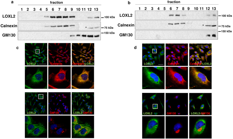
Figure 1. Accumulation of LOXL2 in the ER.
(a,b) Total membrane fractions from MDA-MB-231 (a) and Hs578T (b) cells were fractionated on linear Optiprep gradients and fractions were analysed by immunoblotting using anti-LOXL2 (Origene), and anti-Calnexin and anti-GM130 as ER and cis-Golgi markers, respectively. (*) Unrelated protein. (c,d) Immunofluorescence staining of LOXL2 (green), Calnexin and GM130 (red) in MDA-MB-231 (c) and Hs578T (d) cells; merge images are shown on the right panels. Nuclei were counterstained with DAPI (blue); scale bars, 50 μm. Insets in c and d, indicate amplified areas shown in the bottom panels.

