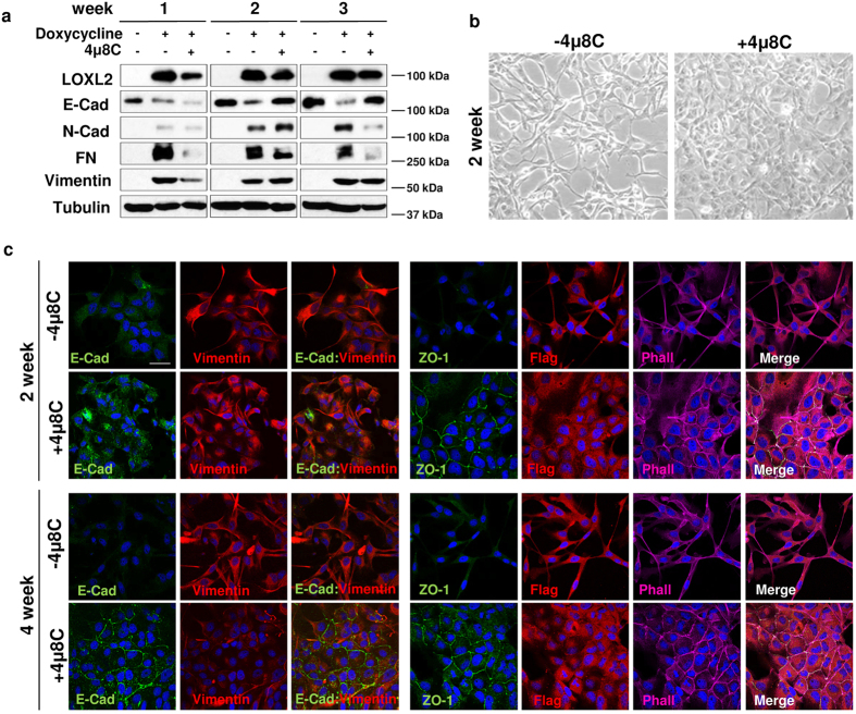
Figure 5. The IRE1-XBP1 branch of UPR mediates the ability of LOXL2 to induce EMT.
MDCK-II cells with inducible expression of LOXL2-Flag were treated with doxycycline and after that with the IRE1 inhibitor 4 μ8 C for the indicated time periods. Cells were processed for: (a) WB using antibodies against LOXL2 (anti-Flag), E-cadherin (E-Cad), N-cadherin (N-cad), Fibronectin (FN) and Vimentin. α-tubulin was used as loading control. One representative blot of two independent experiments is shown. (b) Phase contrast image of the cells after 2 weeks of inhibitor treatment (right) compared to control untreated cells (left). (c) Representative images of confocal immunofluorescence analyses of control and 4 μ8 C treated cells for the indicated time periods with antibodies against LOXL2 (anti-Flag), E-cadherin (E-cad), ZO-1 and vimentin. F-actin was detected with phalloidin stain. Merge images are shown on the right panels. Scale bars 50 μm.

