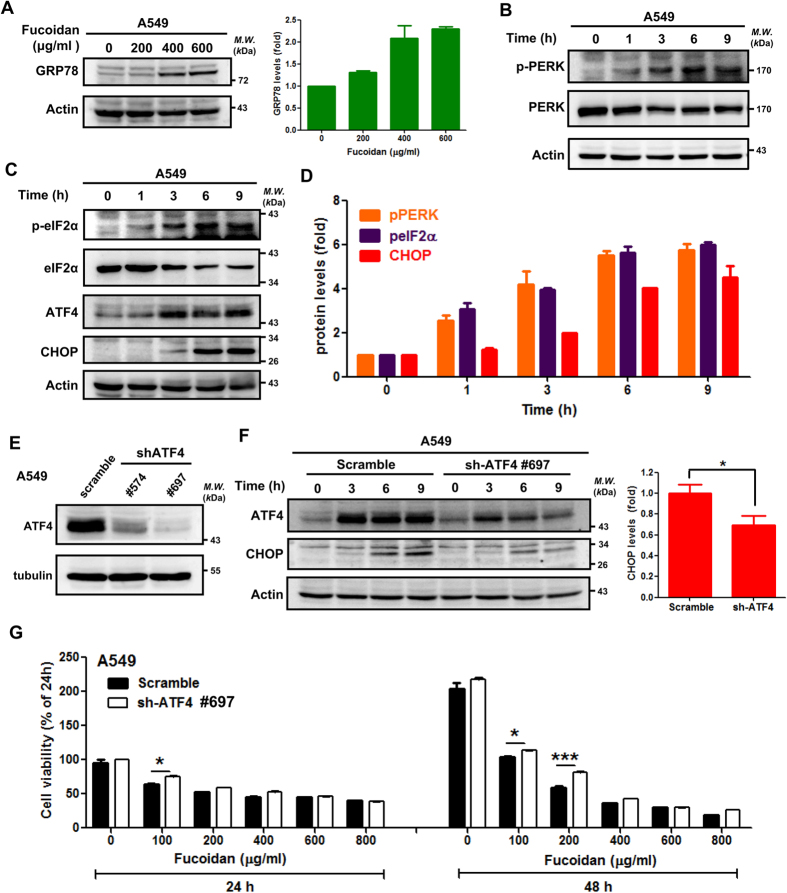
Figure 4. Fucoidan induces ER stress, and ATF4-shRNA (sh-ATF4) interferes with fucoidan-induced CHOP expression in lung cancer cells.
(A) A549 cells were treated with fucoidan (0–600 μg/ml) for 48 h, and Western blotting analyses of whole cell lysates were subsequently performed to detect GRP78 expression. Right panel: Quantification of the intensities of GRP78 bands, which is representative of three separate determinations with ImageJ. (B,C) A549 cells were treated with fucoidan (400 μg/ml) for 1–9 h, and Western blotting analyses of whole-cell lysates were subsequently performed to detect the expressions of ER stress-related proteins. (D) Quantification of the band intensities of pPERK, peIF2α and CHOP (B-C) is representative of three separate determinations with ImageJ. (E) ATF4 expression in A549 cells was detected by Western blotting of whole-cell lysates following TG treatment for 24 h. (F) A549 cells (scramble and ATF4-knockdown) were incubated with fucoidan (200 μg/ml) for 0–9 h, and Western blotting of whole-cell lysates was subsequently performed to detect ATF4 and CHOP. Actin was used as an internal control. Right panel: The densitometric values of CHOP normalized to the relative actin value. Quantification of the intensities of CHOP bands was performed following fucoidan treatment for 9 h. The data are presented as the mean ± the SD, and error bars indicate the SD. Significant differences are noted (*P < 0.05 compared with the control group). (G) A549 cells (scramble and ATF4-knockdown cells) were treated with various doses of fucoidan (μg/ml) for 24 and 48 h. Cell viabilities were determined by crystal violet staining assays. Each group with fucoidan was normalized against each untreated control.

