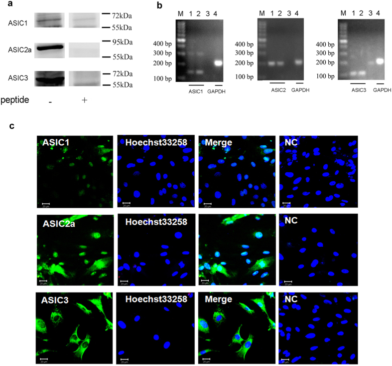
Figure 2. Expressions of ASIC subunits in rat cardiomyocytes.
(a) Expressions of ASIC1, 2a and 3 proteins in rat cultured cardiomyocytes. Individual ASIC subunits peptides were added as negative controls. The blots were cropped from Supplementary Fig. S1a. The representative full-length blot for ASIC2a was shown in Supplementary Fig. S2. (b) Expressions of ASIC1, 2 and 3 transcripts in cultured rat ventricular myocytes. GAPDH transcript was used as control. M: marker; 1, 4: cardiomyocytes; 2: cortex; 3: negative control. (c) Double - labeling fluorescence of ASICs (green) and nucleus (blue, marker: Hoechst 33258) in cultured ratcardiomyocytes. NC: pretreatment with immunogenic peptide as negative control. Scale bars: 20 μm. All data were represented from at least three similar independent experiments.

