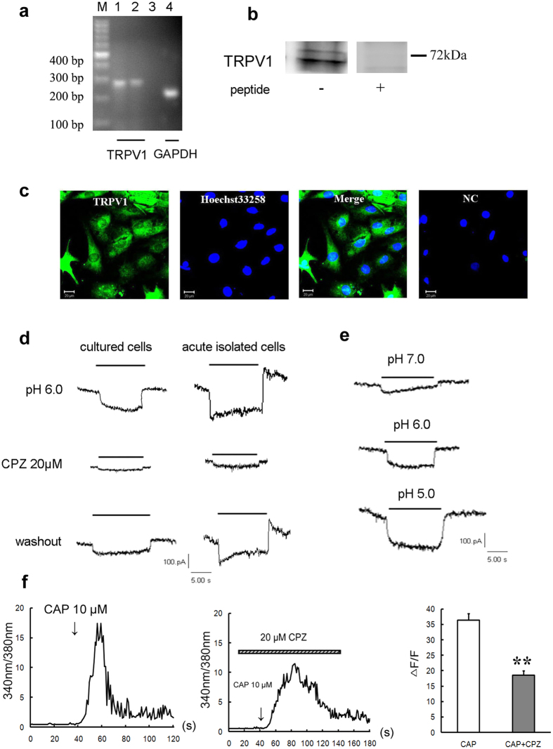
Figure 4. Functional expression of TRPV1 channel in rat cardiomyocytes.
(a) RT-PCR detection of TRPV1 mRNA expressions in cultured rat ventricular cardiocytes. GAPDH were used as positive controls. M: marker; 1, 4: cardiomyocytes; 2: cortex; 3: negative control. (b) Western blotting indicating the protein expression of TRPV1 in cultured ventricular cardiocytes of rat. TRPV1 peptide was used as negative control. The blots were cropped from Supplementary Fig. S1b. (c) Double immunostaining of TRPV1 (green) and nucleus (blue, marker: Hoechst33258) in rat cultured cardiomyocytes. NC: pretreatment with immunogenic peptide as negative control. Scale bars: 20 μm. Above all data were represented from three similar independent experiments. (d) TRPV1-like currents reversibly inhibited by CPZ (20 μM, n = 3 cells) in cultured and acute isolated rat cardiomyocytes. (e) Representative TRPV1 current traces evoked by indicated pH solutions from pH 7.4 in cultured rat cardiomyocytes. “—” indicates the duration of pH = 7.0, 6.0 or 5.0. (f) Representative [Ca2+]i responses and quantitative analysis of normalized Fura-2/AM fluorescence induced by capsaicin (10 μM, n = 16 cells) and CPZ (20 μM) + capsaicin (10 μM, n = 16 cells). Data were shown as mean ± s.e.m. **P < 0.01 vs capsaicin (Student’s t-test). CAP: capsaicin.

