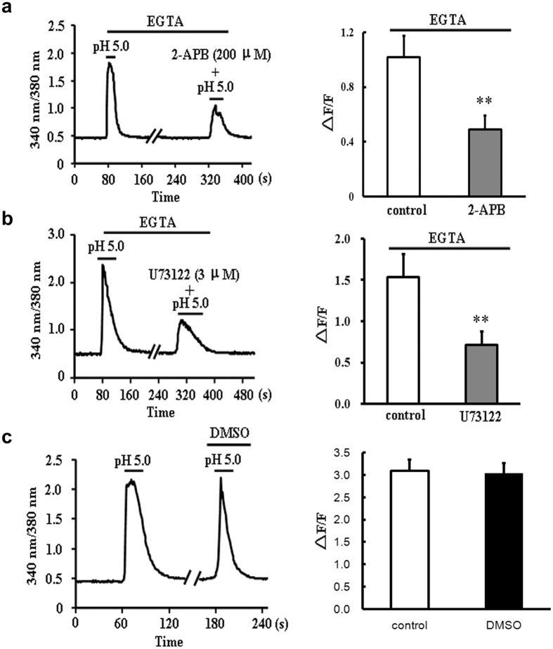
Figure 6. The effects of 2-APB and U73122 on pH 5.0 solution-induced [Ca2+]i elevation in the absence of extracellular Ca2+.
(a) Representative 340/380 nm ratio and summary data (∆F/F) of primary cardiomyocytes showing the changes in [Ca2+]i induced by pH 5.0 solution in the absence or presence of 2-APB (200 μM) (n = 15 cells for each group). Data were shown as mean ± s.e.m (**P < 0.01 vs control, Student’s t-test). (b) Representative 340/380 nm ratio and summary data (∆F/F) of primary cardiomyocytes showing the changes in [Ca2+]i induced by pH 5.0 solution in the absence or presence of U73122 (3 μM) (n = 13 cells for each group). Data were shown as mean ± s.e.m (**P < 0.01 vs control, Student’s t-test). (c) Representative traces of 340/380 nm ratio and summary data (∆F/F) of primary cardiomyocytes showing no changes of [Ca2+]i (pH 5.0) in the presence of 0.1% DMSO (conrol: n = 12 cells, DMSO: n = 18 cells, P > 0.05 vs control, Student’s t-test).

