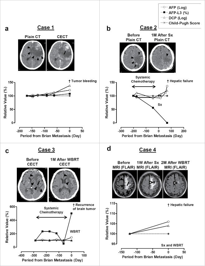
Figure 1.
CT and/or MRI images of brain tumor and clinical courses of Case 1 (a), Case 2 (b), Case 3 (c), and Case 4 (d). CECT, contrast enhanced CT; Sx, surgical resection; WBRT, whole brain radiation therapy. Black arrowheads indicate area of swelling. Black arrows indicate tumors and bleeding from the tumor. White arrowheads indicate the change after Sx and WBRT. AFP and DCP are monitored its increment ratio using their log value and comparing with the initial value available in our hospital. AFP-L3 (%) and Child-Pugh score were monitored for its changes from initial value.

