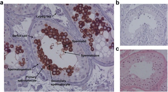
Fig. 3.

Immunohistochemical staining of TEX101 protein in testicular tissue with active spermatogenesis. a Testicular tissue stained with our monoclonal antibody 23ED228 (final concentration 80 ng/mL). Cell types presented include Sertoli cells (negative staining), Leydig cells (negative staining), and germ cells at different stages of spermatogenesis, such as spermatogonia (negative), primary spermatocytes (positive cytoplasm), secondary spermatocytes (positive membrane), spermatids (positive), and spermatozoa. b Negative control (no primary antibody added). c Hematoxylin and eosin staining of testicular tissue showing nucleus (purple) and cytoplasm (pink)
