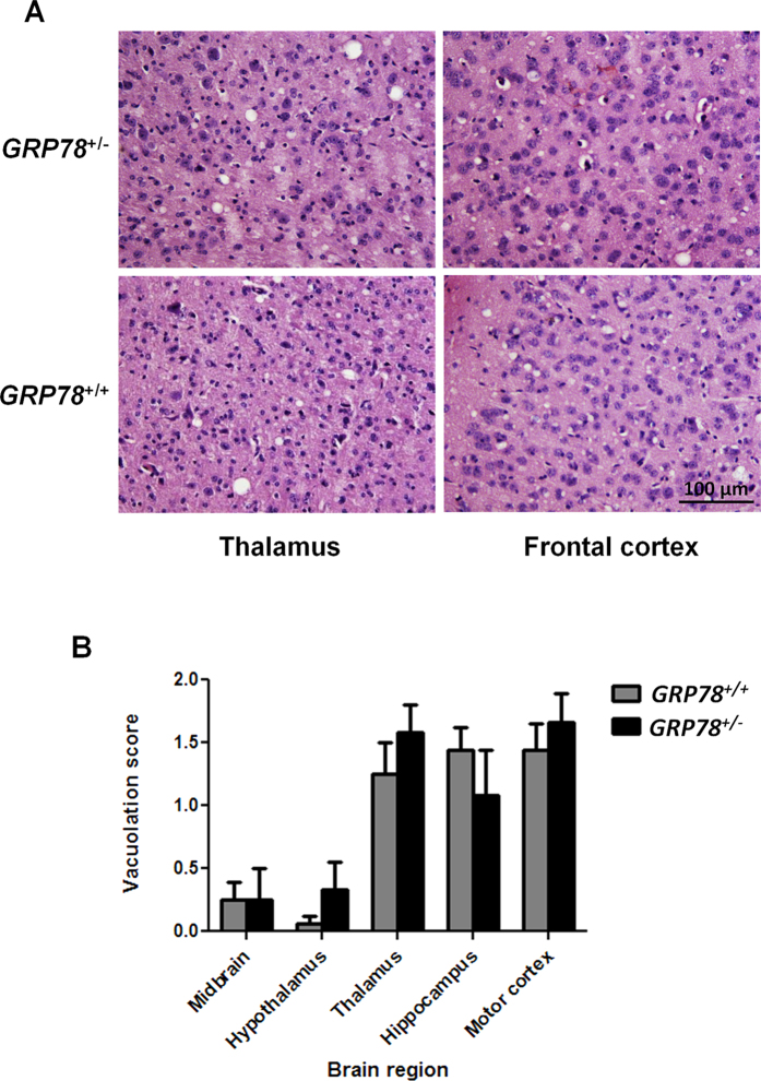
Figure 2. GRP78 expression does not alter the vacuolation profile of terminally ill prion infected mice.
(A) Thalamus and frontal cortex sections of brains from RML-symptomatic GRP78 heterozygous (Grp78+/−) and wild type (Grp78+/+) mice were analyzed histologically for spongiform degeneration after hematoxylin-eosin staining. Bar in the lower right panel depict 100 μm and is representative of all pictures in this panel. (B) The vacuolation lesion profiles were determined on H&E stained sections from 5 different animals in each group. Degree of vacuolation was analyzed by scoring midbrain; hypothalamus; thalamus; hippocampus and motor cortex.

