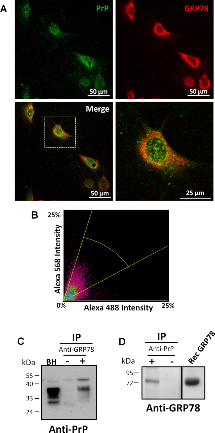
Figure 4. GRP78 interacts with PrP.
(A) Primary cultures of mouse fibroblasts were doubly labeled with antibodies against PrP and GRP78 proteins. Top left panel represents cells that have been labeled with the 6H4 anti-PrP antibody and detected with Alexa488 secondary antibody (green). Top right panel represents cells that have been stained with anti-GRP78/BiP and detected with Alexa568 secondary antibody (red). Bottom left panel represents the merge between the two staining. Bottom right panel is a zoomed picture of one cell of the merged pictures (depicted in the dotted box in the bottom left panel). Samples were visualized by a confocal microscope. Scale bar: 50 μm or 25 μm. (B) Representative fluorogram indicating the signal intensity for both stainings and the colocalization of 6H4 (Alexa 488) and GRP78 (Alexa 568) obtained from confocal images. (C) Wild type mouse brain homogenates were immunoprecipitated with the anti-GRP78 antibody. Samples were analyzed by Western blot using an anti-PrP antibody (6D11). Lane 1 represents untreated brain homogenates used as a control, lane 2 corresponds to precipitation done with uncoated beads (without anti-GRP78 antibody), and lane 3 represents the immunoprecipitation with anti-GRP78 antibody. (D) Wild type mouse brain homogenates were immunoprecipated with the 6D11 anti-PrP antibody and samples analyzed by Western blot with anti-GRP78 antibody. First lane corresponds to the immoprecipitation with the 6D11 antibody, whereas the second line is the precipitation with the beads alone. Third lane depicts recombinant GRP78. Numbers on the left side of the gels correspond to the molecular weight standards. Separation line in the right blot indicate gel splicing to remove some irrelevant lines, even though all the samples were run in the same gel.

