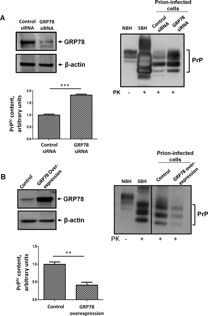
Figure 5. Expression levels of GRP78 modify prion replication in chronically infected CAD5 cells.
(A) Prion-infected CAD5 cells transfected with GRP78 siRNA or control siRNA were harvested and lysed. The expression of GRP78, actin (loading control), and PrPSc was analyzed by Western blotting. Left blot shows the staining with GRP78 antibody. Right blot corresponds to the staining with anti-PrP antibody. The graph shows the densitometric analysis of the levels of PrPSc in cells treated with control or GRP78 siRNA. (B) Prion-infected CAD5 cells transfected with GRP78 overexpressing plasmid or control plasmid were harvested and lysed. The expression of GRP78, actin, and PrPSc was analyzed by Western blotting. Left blot depicts the staining for GRP78. Right blot corresponds to the staining for PrP. In this panel, the vertical line indicates gel splicing to remove some irrelevant lanes, but samples were run in the same gel and were developed with the same exposition. The graph shows the densitometric analysis of PrPSc levels in cells expressing endogenous amounts of GRP78 (control) or over-expressing this protein. In both panels A and B, NBH: normal brain homogenate, not treated with PK, used as a marker of PrPC migration. SBH: RML-infected brain homogenate treated with PK, used as a marker of protease-resistant PrPSc migration. For space constrains some blots were cropped, but all samples were run using the same conditions and in the same gel. Statistical differences were analyzed by using student’s t-test. **P < 0.01, ***P < 0.001.

