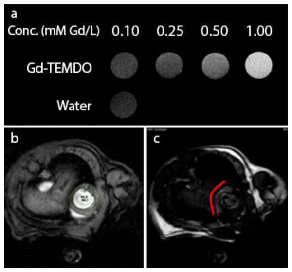
Figure 2.

Gd-TEMDO MRI. (a) T1-weighted MRI phantoms of Gd-TEMDO proving concentration dependent T1 shortening. (b) MRI obtained from isoflurane-anaesthetized mice, (c) taken 30 minutes after I.P. administration of Gd-TEMDO (0.6 mmol/kg). Left: the heart fully visible; right: heart with reduced brightness, the damaged tissue remains visible due to absorbed Gd-TEMDO following the red line.
