
Figure 2.
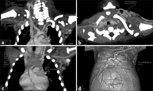
Contrast-enhanced computed tomography scan shows collaterals due to anomalies of the left brachiocephalic and subclavian veins and hypoplastic right internal jugular vein. (a) Venous collaterals in the right upper posterior area of the neck draining into the lower internal jugular vein that is hypoplastic. (b) The left brachiocephalic vein is not seen. (c) Venous collaterals around the left clavicle. (d) Surface collaterals as shown in a three-dimensional computed tomography image