
Figure 1.
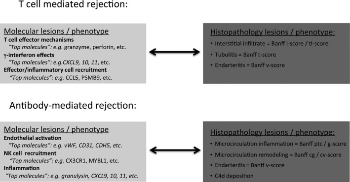
Molecular lesions and their corresponding histologic lesions in T cell–mediated rejection and antibody‐mediated rejection in kidney allografts. cg, glomerular double contours; cv, vascular fibrous intimal thickening; i, inflammation; ptc, peritubular capillaritis; ti, total inflammation; v, intimal arteritis.