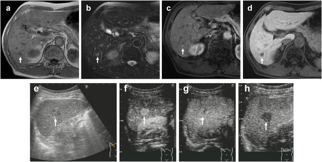
Figure 1.

A 70‐year‐old man with a history of nonalcoholic steatohepatitis underwent surgical resection of transverse colon cancer. Follow‐up imaging showed a 1.7‐cm tumor in S6. (A) Gd‐EOB‐DTPA‐enhanced MRI (EOB‐MRI) shows a hypointense lesion in T1‐weighted images (arrow). (B) EOB‐MRI shows a hyperintense lesion in T2‐weighted images (arrow). (C) EOB‐MRI shows a hyperintense lesion in arterial phase (arrow). (D) EOB‐MRI shows a hypointense lesion in hepatobiliary phase (arrow). The lesion had a diagnosis of liver metastasis (confidence scale 4). (E) Gray‐scale US shows a hypoechoic lesion in S6 (arrow). (F) Contrast‐enhanced US (CEUS) in arterial phase (40 seconds) shows hyperenhancing lesion (arrow). (G) CEUS shows isoenhancing lesion in late phase (arrow). The enhancement lasted more than 120 seconds. (H) CEUS shows hypoenhancing lesion in postvascular phase (arrow). The lesion had a diagnosis of not liver metastasis (confidence scale 1) from diagnostic criteria of CEUS, which was diagnosed hepatocellular carcinoma histopathologically.
