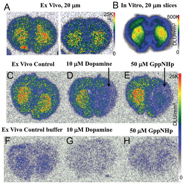
FIGURE 6.
Ex Vivo 18F-5-OH-FHXPAT Studies: (a) Rats injected with ~7 MBq of 18F-5-OH-FHXPAT and killed at 25 min postinjection. Coronal brain slices (20 μm) were obtained showing greater binding in striatum (ST), with striata to cortex ratio of 1.5–1.6. (b) In vitro binding observed in rat brain coronal sections incubated with 18F-5-OH-FHXPAT at 37°C for 30 min. (c–e) Ex vivo coronal sections showing total binding at 25 min post injection (c), 10 μM dopamine applied for 5 min to the right half displacing 18F-5-OH-FHXPAT from the striata (d) and 50 μM Gpp(NH)p applied for 5 min to the right half displacing 18F-5-OH-FHXPAT from the striata (e). (f–h) Ex vivo coronal sections obtained 25 min postinjection showing 12 min buffer-treated total binding (f), buffer containing 10 μM dopamine treatment reduced binding by >80% (g) and buffer containing 50 μM Gpp(NH)p treatment, which reduced binding by >45% (h)

