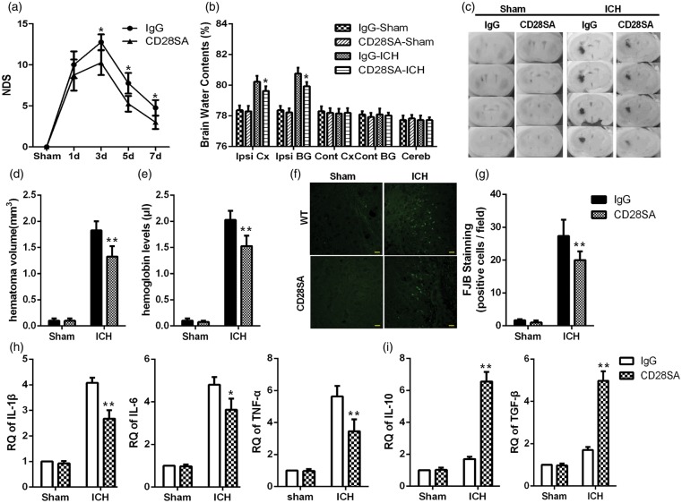
Figure 4.
CD28-SA ameliorated inflammatory injury after ICH. (a) The NDSs in the IgG and CD28-SA groups at 1, 3, 5, and 7 d after ICH (*p < 0.05 versus the IgG group at the corresponding time points, n = 12 for each time point). (b) Brain water content at 4 d after hemorrhage or sham operation. (c) Serial coronal sections of mouse brain tissues at 4 d after ICH. (d) Brain sections were used to measure the hematoma volume. (e) Brain homogenate was used to measure the hemoglobin level (**p < 0.01, *p < 0.05 versus the WT-ICH group, n = 4). (f) Representative FJB-positive cells in the CD28-SA and IgG groups (bars=20 µm). (g) FJB-positive cells were quantified and compared (**p < 0.01 versus IgG, n = 6). (h) Global changes in M1-related inflammatory factors. (i) Global changes in M2-related inflammatory factors at 4 d after hemorrhage or sham operation were analyzed using RT-PCR (*p < 0.05, **p < 0.01 versus the IgG group, n = 4).

