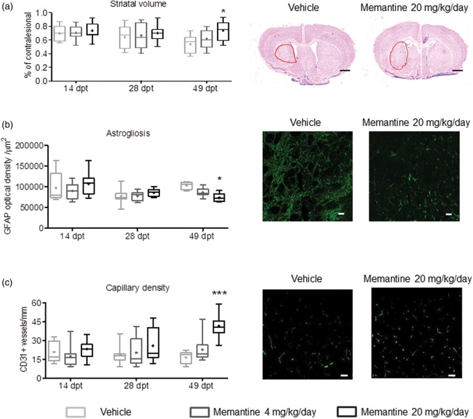
Figure 4.
Memantine promotes peri-infarct brain remodeling. (a) Striatal volume, evaluated on cresyl violet-stained brains sections, (b) diffuse astrogliosis, evaluated by GFAP immunohistochemistry, and (c) capillary density, evaluated by CD31 immunohistochemistry, in animals exposed to transient MCAO that were subcutaneously treated with vehicle or memantine (4 or 20 mg/kg/d) starting 72 h after reperfusion onset. Photomicrographs at 49 dpt are also shown. Optical density in (b) was evaluated in the peri-infarct (parietal) cortex, whereas capillary density in (c) was assessed in a total of six ROI within the striatum, as described in the Materials and methods section. Representative microphotographs from these brain regions are shown. Results are medians (lines inside boxes)/means (crosses inside boxes) ± IQR (boxes) and minimum/maximum data (elongation lines) (n = 12 animals/ group). Data were analyzed by two-way ANOVA followed by two-tailed unpaired t tests for individual time-points. *p < 0.05, ***p < 0.001 compared with ischemic vehicle. Bars, 1000 µm (a)/25 µm (b and c).

