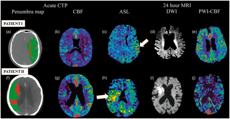
Figure 1.
Multimodal imaging (acute CTP and follow-up 24 h MRI) showing perilesional hyperperfusion (PLH) on ASL at 24 h. Patient I (upper row): CTP demonstrated acute left MCA territory infarct with a small core and large penumbra (a), and decreased rCBF (b). Post-thrombolysis, patient showed excellent recanalisation. PLH patterns were observed on ASL-MRI at 24 h (c). Area of restricted diffusion was observed on follow-up DWI-MRI (d). PWI-CBF at 24 h is also depicted (e). Patient II (bottom row): Acute CTP shows large penumbra (f) and decreased rCBF (g) over a large part of the right MCA territory indicating a large area of hypoperfused but likely viable tissue (penumbra). Post-thrombolysis, the patient showed excellent response to therapy. The regions that were previously hypoperfused showed PLH (white arrow) at 24 h on ASL (h). DWI-MRI (i) demonstrated an area of restricted diffusion. Hyperperfusion patterns are not clearly evident on PWI-CBF (j). Both patients showed good collaterals on baseline CTA.
ASL: arterial spin labelling; CBF: cerebral blood flow; CT: computed tomography; CTA: computed tomographic angiography; CTP: computed tomographic perfusion imaging; DWI: diffusion-weighted imaging; MCA: middle cerebral artery; MIP: maximum intensity projection; PWI: perfusion-weighted imaging.

