Figure 7.
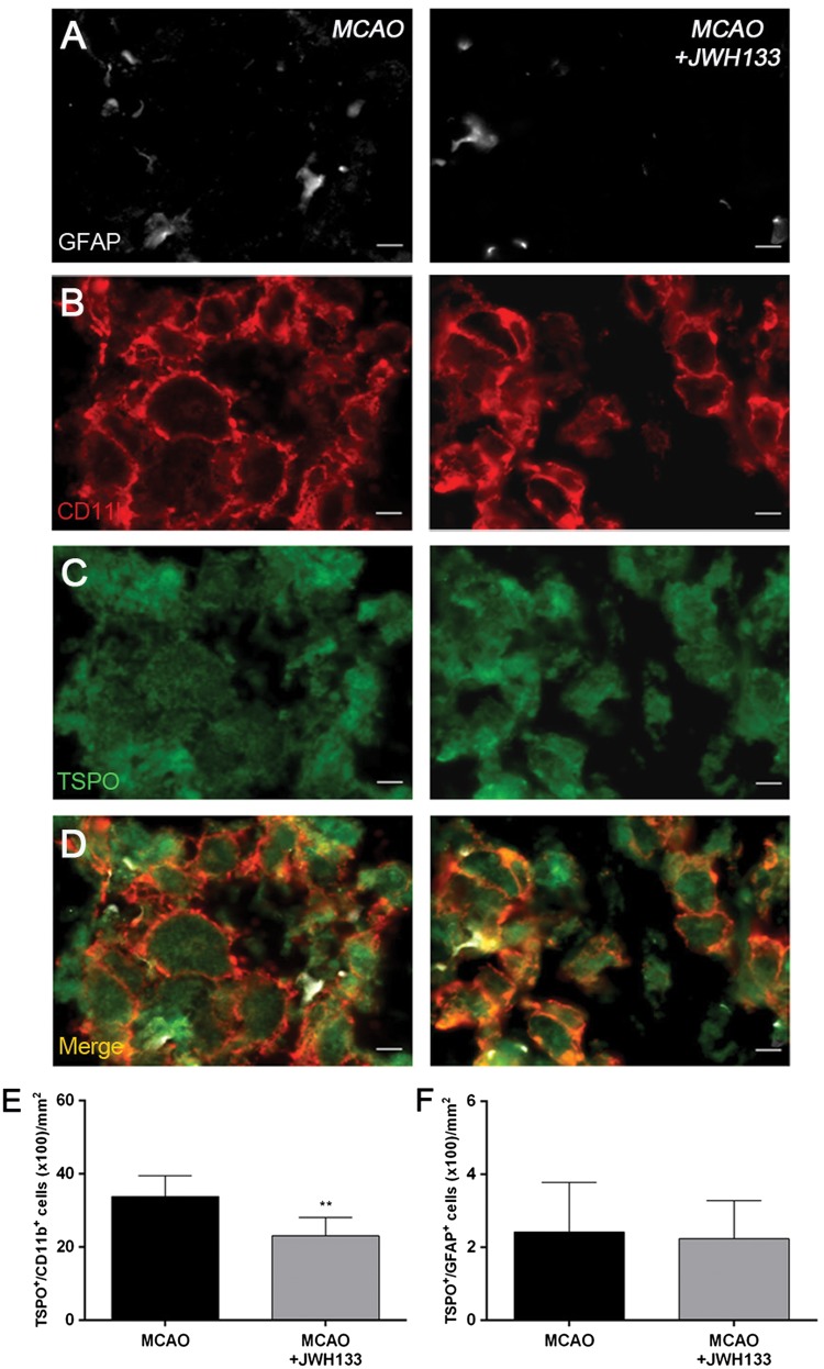
Immunofluorescent labeling of GFAP (white), CD11b (red) and TSPO (green) in the ischemic area, shown as three channels. The data show temporal evolution of TSPO in microglial and astrocytic cells at day 7 after MCAO in vehicle (left column, n = 5) and JWH133-treated rats (right column, n = 7). GFAP-positive astrocytes do not change after treatments (a). CD11b-reactive microglia/macrophages (b) and TSPO receptor (c) decrease after MCAO in JWH133-treated rats. (d) Merged images of three immunofluorescent antibodies. The number of CD11b-reactive microglia/macrophages expressing TSPO decrease at day 7 after daily treatment with JWH133 (e). The number of GFAP-reactive astrocytes expressing TSPO shows similar values following treatment (f). **p < 0.01 compared with vehicle. Scale bars, 5 µm.

