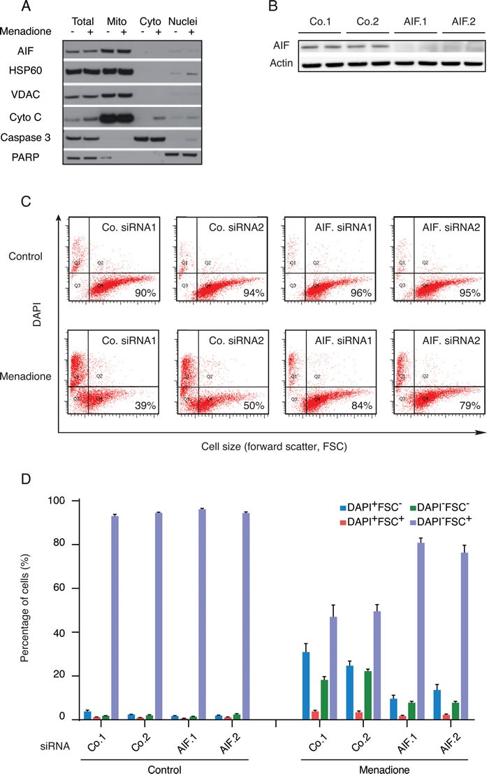
Figure 1. Impact of AIF expression on the cytotoxicity of menadione.

A. After 3h treatment with (+) or without (−) 50μM menadione, the subcellular localization of AIF in U2OS cells was checked by immunoblot in the indicated fractions, using antibodies directed against proteins localized in mitochondria (HSP60, VDAC and cytochrome c), cytosol (caspase-3) or nuclei (PARP). B. Extracts of U2OS cells (duplicates) subjected to the transfection with AIF-specific (AIF.1 and AIF.2) or control (Co.1 and Co.2) siRNA were analyzed by immunoblot for the abundance of AIF. Actin was used as a loading control. C. D. U2OS cells transfected with two distinct control siRNAs (Co.1 and Co.2) or two distinct, non-overlapping siRNAs targeting AIF (siRNA AIF.1 and AIF.2) were treated with 50μM menadione or the solvent (Control) for 3h and drug-induced cell death was quantified by flow cytometric assessment (pictograms shown in C; histograms shown in D) of DAPI uptake (DAPI positivity) and forward light scatter (FSC) analysis that allows for the identification of apoptotic cells according to their reduced size (low FSC). Data are expressed as mean values ± SEM.
