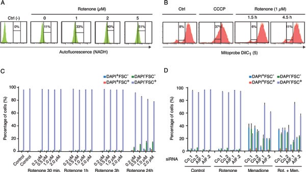
Figure 3. Effect of respiratory chain complex I inhibition on Menadione-induced cytotoxicity.

A. Dose-dependent rotenone-induced respiratory chain complex I inhibition was monitored by flow cytometry, after excitation with a UV laser. The augmentation of cell fluorescence reflects the accumulation of NADH in the treated cells. The NADH-dependent autofluorescence of cells incubated with 1 to 5 μM of rotenone for 4.5 h was compared to that of untreated cells (0 μM) and negative control cells (Ctrl) permeabilized with 70% ETOH before analysis. B. The mitochondrial membrane potential of cells incubated with 1 μM rotenone for 1.5 h or 4.5 h was monitored by flow cytometry, using the MitoProbe Dil C1(5), and compared to that of control cells untreated (Ctrl) or treated with the protonophore CCCP, which dissipates the mitochondrial transmembrane potential. For each condition, the percentage of cells showing a reduced Dil C1(5) incorporation is mentioned. C. The effect of rotenone on cell survival was quantified by flow cytometric assessment of DAPI uptake (DAPI positivity) and forward light scatter (FSC) analysis, after incubation with 0 to 2μM of Rotenone for the indicated times. D. Effects of AIF knockdown, combined or not with rotenone treatment (1 μM for 4.5 h), on menadione-induced (50 μM for 3 h) cytotoxicity was analyzed after transfection with two distinct control siRNAs (Co.1 and Co.2) or two distinct, non-overlapping siRNAs targeting AIF (siRNA AIF.1 and AIF.2). Cell death was monitored by flow cytometric assessment of DAPI uptake (DAPI positivity) and forward light scatter (FSC) analysis. Data are expressed as mean values ± SD.
