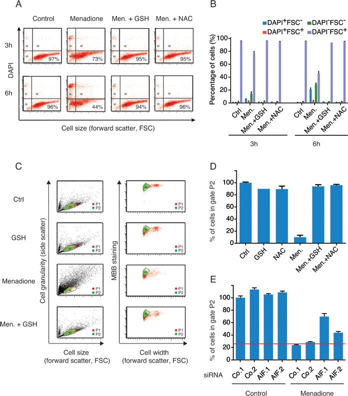
Figure 4. The loss of GSH levels in menadione-treated cells correlates with the expression level of AIF.

A., B. Effect of exogenous antioxidants on menadione-induced death was evaluated by incubating U2OS cells, for 3h or 6h, with 50μM of menadione in the absence or presence of GSH (5 mM) or NAC (5 mM). Cell death was quantified by flow cytometric assessment (pictograms are shown in A and histograms in B) of DAPI uptake (DAPI positivity) and forward light scatter (FSC) analysis that allows the identification of apoptotic cells. C., D. A cytofluorimetric analysis combined with the use of the thiol-reactive probe monobromobimane (MBB) was set up to measure levels of reduced glutathione in cells treated with menadione (pictograms are shown in C and histograms D). After menadione treatment, in absence or presence of exogenous antioxidants (GSH or NAC), live cells (Topro3 negative), exhibiting size and granularity parameters similar to control untreated cells (gate P1), were analyzed for their staining with MBB (gate P2). Cell width assessment by forward light scatter (FSC) analysis was used to discriminate between singlet cells and aggregates. For each treatment condition, the percentage of cells stained with MBB (gate P2) was quantified (D). E. The effect of AIF knockdown on the levels of GSH was monitored, as described in (C and D), after transfection with two distinct control siRNAs (Co.1 and Co.2) or two distinct, non-overlapping siRNAs targeting AIF (siRNA AIF.1 and AIF.2) and culture with 50 μM of menadione for 3h. Data are expressed as mean values ± SD.
