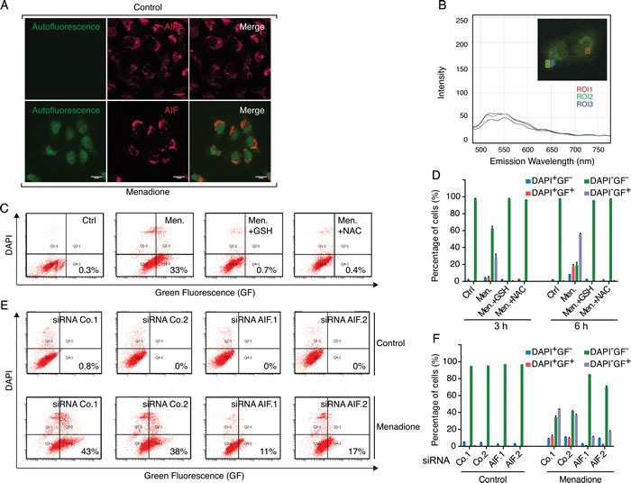
Figure 5. The metabolization of fluorescent menadione-cysteinyl group conjugates correlates with AIF expression levels.

A. Microscopic analysis of U2OS cells revealed that, compared to control conditions (cells treated with the solvent), the incubation with 50 μM menadione for 3 h provoked the appearance of a diffuse cellular fluorescence that resisted to the fixation/permeabilization protocol. The mitochondrial localization of AIF, both in control and menadione-treated cells, was revealed by indirect immunofluorescence, using an anti-AIF rabbit polyclonal antibody and an Alexafluor 647-conjugated secondary anti-rabbit antibody (AIF red staining). Individual and merged images show that in menadione-treated cells, AIF is not released from the mitochondrion and the diffuse distribution of menadione-induced autofluorescence is maximal in the nuclear compartment. B. Emission spectra and intensity analyses of the fluorescence produced in menadione-treated cells were evaluated by microscopy. The insert corresponds to the menadione-treated cell that was imaged by fluorescence microscopy (Zeiss) and squares on the image correspond to distinct regions of interest (ROI1 to to ROI3) that were evaluated for fluorescence spectra. C. D. The formation of fluorescent menadione-cysteinyl group conjugates (green fluorescence, GF) was monitored by flow cytometric analysis of U2OS cells incubated for 3h or 6h with 50 μM menadione, in the absence or presence of exogenous antioxidants GSH (5 mM) or NAC (5 mM). Analyses of the pictograms (C) and histograms (D) reveal that treatments with both exogenous GSH and NAC inhibit the formation of the fluorescent menadione-cysteinyl group conjugates in menadione-treated cells. E. F. After transfection with two distinct control siRNAs (Co.1 and Co.2) or two distinct, non-overlapping siRNAs targeting AIF (siRNA AIF.1 and AIF.2), cells were submitted to menadione treatment (50μM) for 3 h and then analyzed, as in C and D, for the formation of fluorescent menadione-cysteinyl group conjugates. Analyses of the pictograms (E) and histograms (F) reveal that the depletion of AIF inhibits the formation of the fluorescent menadione-cysteinyl group conjugates in menadione-treated cells. DAPI staining was used for monitoring of death-induced membrane permeabilization. Data are expressed as mean values ± SD.
