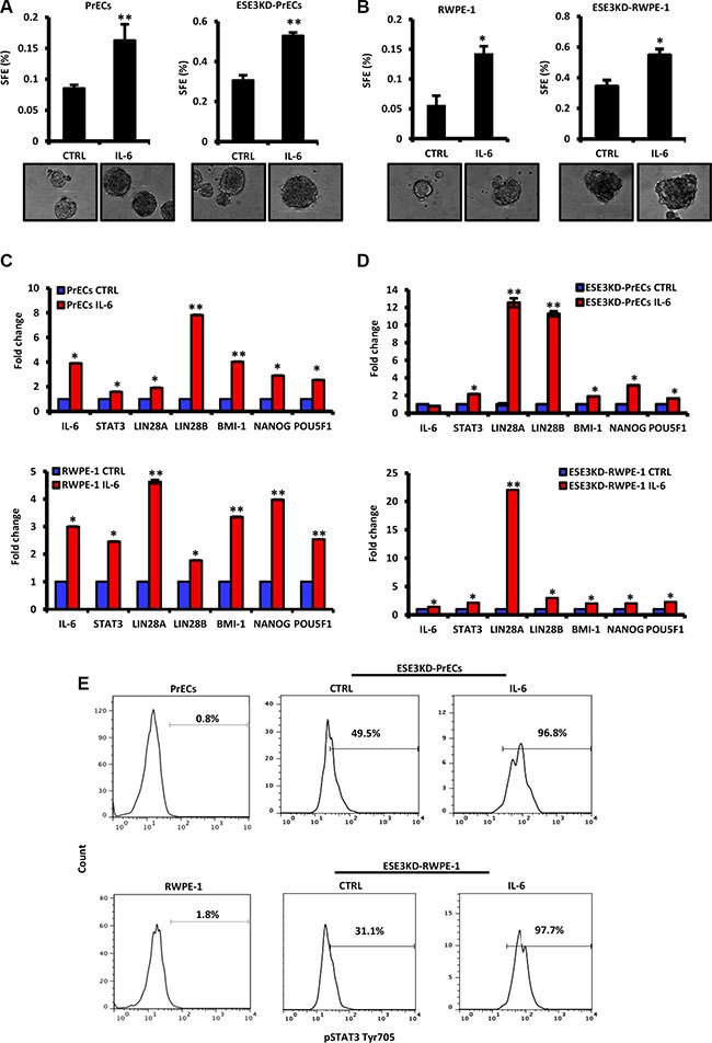
Figure 4. IL-6 induces transformation and cancer stem-like phenotypes in normal and more significantly in ESE3KD prostate epithelial cells.

(A–B) Sphere forming efficiency (SFE) in PrECs and ESE3KD-PrECs (A) and in RWPE-1 and ESE3KD-RWPE-1 (B) cells after IL-6 treatment in vitro. (C–D) mRNA levels of STAT3, IL-6 and indicated genes evaluated by qRT-PCR in PrECs and ESE3KD-PrECs (left) (C) and in RWPE-1 and ESE3KD-RWPE-1 cells (right) (D) following 4 hours of treatments in vitro with IL-6 (50 ng/ml) or DMSO as control (CTRL). Data are presented as fold change relative to control (CTRL) cells. (E) Flow cytometry analysis of STAT3-Tyr705 staining following IL-6 treatment of control PrECs and ESE3KD-PrECs (upper) and RWPE-1 and ESE3KD-RWPE-1 (lower) cells. Percentages of positive cells are shown. P values were determined using t-test. *P < 0.05; **P < 0.01. Data are representative of three independent experiments.
