
Figure 1. Identification of AT deficiency due to a small deletion.
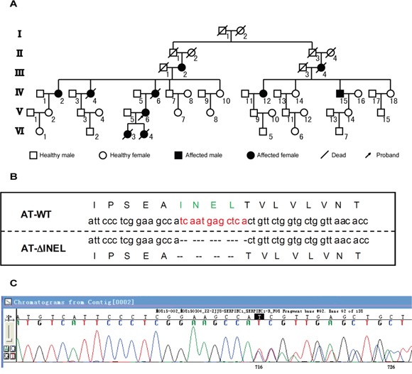
A. Pedigree of proband. The proband is indicated by the arrow. Family members affected with thrombosis are indicated in black, and III-2, IV-2/12/15, and V-6 are the small deletion carriers. B. Representation of the residues deleted (ΔINEL) in the mutant AT aligned with the wild-type (WT) sequence. C. The sequencing graph for mutant AT.