
Figure 1. Detection of CSMD1 mRNA in normal breast tissue and quantitation of CSMD1 mRNA transcript in breast cancer tissues.
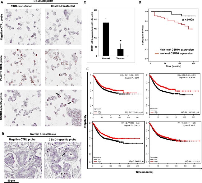
CSMD1-specific probe, as well as a negative (DapB) and a positive (PPIB) control probes were included when staining BT-20 expressing CSMD1 and CTRL paraffin-embedded cell pellets for validation of the method (A). RNAscope detection of CSMD1 mRNA transcripts in paraffin-embedded normal breast tissue. Samples were hybridised with either CSMD1-specific probe or negative control probe. A positive signal for CSMD1 was observed in the normal breast tissues. The black arrows outlined the mRNA brown dots (B). Breast tumor tissues had significantly lower levels of CSMD1 mRNA transcript compared with normal tissues; *p < 0.05 by Mann-Whitney test (C). Patients with low levels of the CSMD1 transcript showed a significantly shorter overall survival (log rank test) (D). Kaplan–Meier plots using as using recurrence-free survival as an endpoint for four probes of CSMD1; HR, hazard ratio (E).