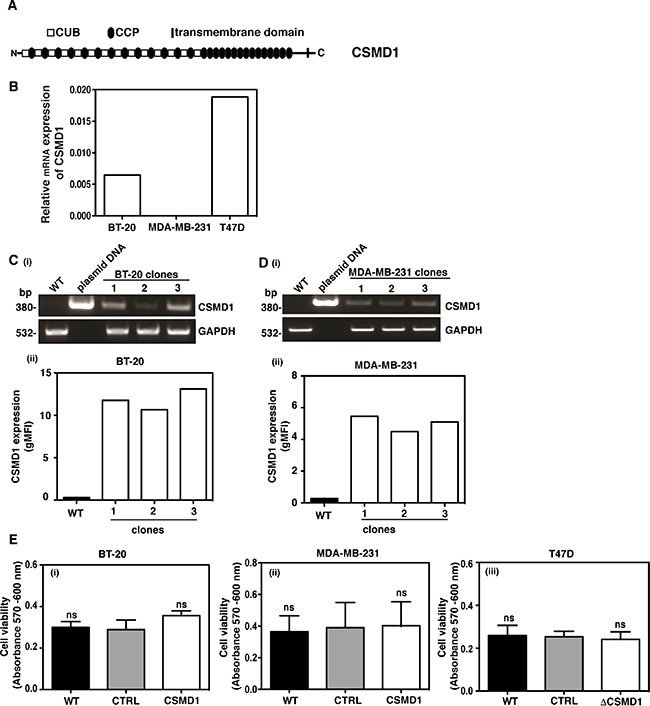
Figure 2. Expression of CSMD1 in breast cancer cell lines.

CSMD1 is composed of CUB and CCP domains followed by a single transmembrane domain and a small cytoplasmic region (A). Screening of breast cancer cell lines for CSMD1 coding sequence at mRNA level using qPCR. The breast cancer cells BT-20 and MDA-MB-231 were selected for expressing CSMD1 and T47D for knocking-down (B). Verification of CSMD1 expression in the 1/2/3 clones for BT-20 and 1/2/3 clones for MDA-MB-231 cells by conventional PCR (i) and flow cytometry (ii). CSMD1 levels were higher when compared to the WT. The data presented is representative of a single experiment performed in duplicates (C–D). The housekeeping gene GAPDH was used as a control of RNA integrity. Cell viability measured after 24h incubation (E) was not affected when expressing CSMD1 in the breast cancer cells BT-20 and MDA-MB-231 (i-ii) or when silencing CSMD1 in the breast cancer cell T47D (iii). Values are means ± SD from 3 independent experiments performed in five replicates. One-way ANOVA was used to calculate statistical significance; ns, not significant.
