
Figure 3. Alteration of CSMD1 expression affects wound healing and migration.
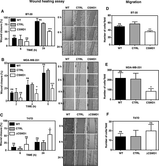
A monolayer of cells was wounded and photographs were taken at different time points. The wound closure was expressed as percentage of wound closure as compared with the zero time point. Percentage wound closure observed in BT-20 (A), MDA-MB-231 (B) and T47D cells (C). WT, CSMD1 or ΔCSMD1 cells were compared to CTRL cells by two-way ANOVA to calculate statistical significance. Motile cells that passed through the pores and adhered to the underside of the cell culture insert membrane following a FBS gradient were photographed and counted for BT-20 (D), MDA-MB-231 (E) and T47D cells (F). A one-way ANOVA was used to calculate statistical significance between the CTRL cells and WT, CSMD1 or ΔCSMD1 cells; *p < 0.05; **p < 0.01; ***p < 0.001; ns, not significant. Results shown are mean of cells counts ± SD from 3 independent experiments performed in duplicates.