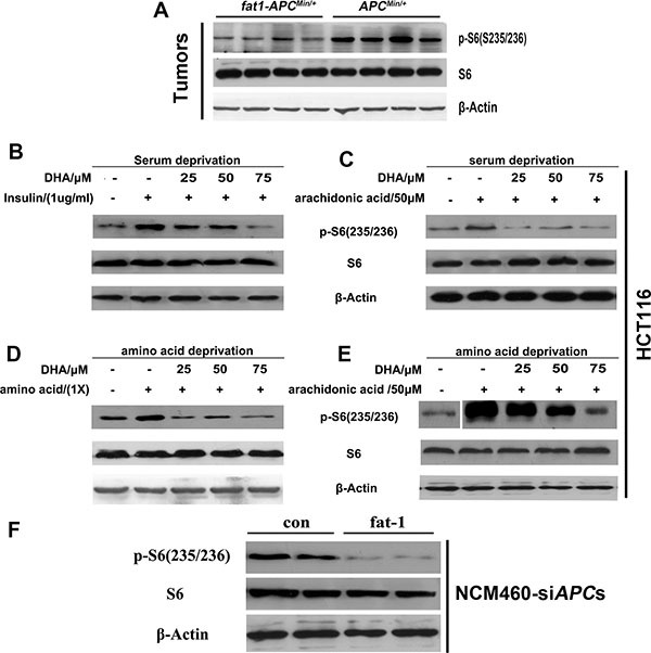
Figure 5. n-3 PUFAs inhibit mTORC1 signaling in vivo and in vitro.

(A) Tumor lysates were subjected to western blotting for measurement of the levels of phosphorylation of S6 (S235/236). HCT116 cells were serum-starved for 18 h, cultured in the presence of the indicated concentrations of DHA for 30 min, and stimulated with insulin (1 mg/ml) (B) or arachidonic acid (50 μM) (C) for 30 min. HCT116 cells were deprived of amino acids and incubated for 30 min in the presence of DHA, followed by incubation with amino acids (1×) (D) or arachidonic acid (50 μM) (E) for 30 min. The protein extract from each set was subjected to western blotting for the analysis of mTORC1 signaling. (F) NCM460 cells previously transfected with APC siRNA were transfected with a pST180 empty vector (con) or with the pST-180-fat-1 vector (fat-1) and serum-starved for 18 h. The protein extract from each set was subjected to western blotting for the analysis of mTORC1 signaling.
