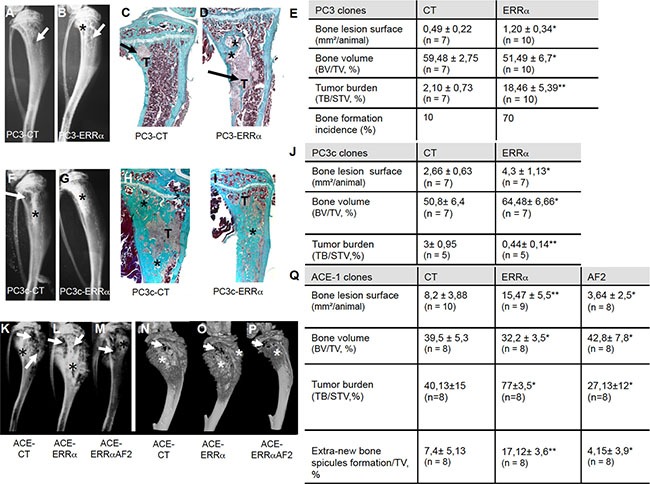
Figure 2. Over-expression of ERRα in prostate cancer cells induced bone lesions development.

Radiography revealed larger lesions in mice injected with (A, B) PC3-ERRα versus PC3-CT, and (F, G) with PC3c-ERRα versus PC3c-CT. Histology after Goldner's trichrome staining confirmed the radiography results in mice injected with (C, D) PC3-ERRα versus PC3-CT (H, I) with PC3c-ERRα versus PC3c-CT (bone matrix in green). (E) Induction -of larger bone lesions surface in mice injected with PC3-ERRα (ERRα)(Mann-Whitney, P = 0.011), -of a decrease in %BV/TV (Mann-Whitney, P = 0.022) and an increase of %TB/STV (Mann-Whitney, P = 0.008) compared with mice injected with PC3-CT (CT). Bone formation incidence show that 70% of mice injected with PC3-ERRα (ERRα) developed some bone formation as opposed to 10% of mice injected with PC3-CT (CT). (J) Increased -of bone lesions surface in mice injected with PC3c-ERRα (Mann-Whitney, P = 0.0175), -of the %BV/TV (Mann-Whitney, P = 0.022) and decrease of the %TB/STV (Mann-Whitney, P = 0.0023) compared with mice injected with PC3c-CT (CT). Radiography (K–M) and 3D micro-tomography reconstructions (N–P) showed larger bone lesions in mice injected with ACE-ERRα versus ACE-CT with an abrogation of the bone lesion effects seen with ERRα overexpression in tumors bearing the dominant negative AF2-truncated ERRα. (Q) After 3 weeks post inoculation of ACE-ERRα, ACE-CT and ACE-AF2 cells, radiography revealed larger and smaller bone lesions surface in mice injected with ACE-ERRα and ACE-AF2 respectively compared to CT (Mann-Whitney, P = 0.0079, P = 0.0304) and microtomographic reconstructions of tibiae show a decrease in mice injected with ACE-ERRα compared to CT (%BV/TV: Mann-Whitney, P = 0.0411), an increase in %TB/STV (Wilcoxon, P = 0.034) compared to CT, and an increase in % new bone formation/TV (extra-bone spicules formation): (Mann-Whitney, P = 0.0025). The increase in bone lesion surface (Mann-Whitney, P = 0.0011), in the %TB/STV (Mann-Whitney, P = 0.0052), in the extra-bone spicules formation (Mann-Whitney, P = 0.0012) and the decrease in %BV/TV (Wilcoxon, P = 0.0273) effects seen with ERRα overexpression were markedly abrogated in tumors bearing the dominant negative AF2-truncated ERRα. * = P < 0.05; ** = P < 0.001; *** = P < 0.0001. T: Tumor; * bone formation; Arrow: bone degradation.
