
Figure 5. Stimulation of VEGF-A expression in PCa cells by ERRα.
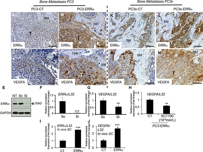
(A–B) Visualization of the overexpression of ERRα protein expression in tumor by IHC on bone lesions in vivo induced by PC3-ERRα (ERRα)(B) compared PC3-CT (CT)(A). (C–D) Paralleling the overexpression of ERRα, VEGF-A expression in tumor was also stimulated in vivo in bone lesions induced by PC3-ERRα (ERRα) (D) compared PC3-CT (CT) cells (C). (E) Decreased ERRα protein expression by transfection of three pooled siRNA sequences in parental PC3 cells shown by Western blot and (F–G) by real-time RT-PCR for ERRα and VEGF-A expression (Student's t-tests P = 0.0002; P = 0.0027). (H) Decreased VEGF-A mRNA expression was also observed after XCT-790 treatment at 10−6M for 48 h in PC3-ERRα cells (Student's t-tests P = 0.006). (I–J) Real-time RT-PCR was performed on triplicate samples and normalized against the ribosomal protein gene L32 to evaluate ERRα (P = 0.0001) and VEGF-A (P = 0.0001) expression within subcutaneous (SC) tumors in vivo (pool of n = 3 for each condition). (K–N) Similarly, to the PC3 model, IHC revealed that ERRα and VEGF-A protein levels in tumors were increased in mixed lesions induced by PC3c-ERRα (ERRα) (L and N concomitantly) compared to PC3c-CT (CT) (K and M concomitantly) in vivo. Bar = 200 μm, T: Tumor; OB: osteoblasts; BM: Bone Matrix.