
Figure 1. Effect of PDGFR-alpha overexpression on endothelial (HUVEC) and melanoma (SKMel-28) cells proliferation.
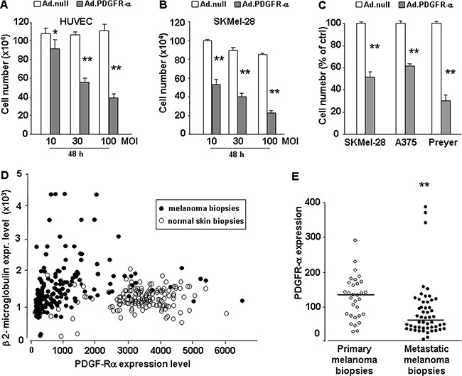
Proliferation of HUVEC and SKMel-28 infected with Ad-vector codingfor PDGFR-alpha. A, B.: Dose-dependent effects of 10, 30, and 100 MOI AdCMV.PDGFR-alpha infection; 10% FCS–induced proliferation at 48 hours. *p value < 0.05 and **p value <0.01 versus AdCMV.null (cell number at T = 0 corresponds to 7.5 ×10⁵). Data are reported as mean ± SD of 3 independent experiments. C. Effects of AdCMV.PDGFR-alpha infection in SKMel-28, A375 and Preyer cell lines. Data are reported as % of control D. Expression of PDGFR-alpha (X axis) vs expression of the housekeeping gene beta 2-microglobulin (Y axis) in 208 melanoma biopsies (black spots) and 147 normal skin biopsies. Expression data were derived from ist.medisapiens.com site. Supplementary Figure 2SA-2SB reports correlation plots of PDGFR-alpha with other housekeeping genes, namely Tubulin Beta 1 gene (Supplementary Figure 2SA) and Actin beta gene (Figure 2SB). In all cases PDGFR-alpha is strongly reduced in melanoma samples vs normal skin samples. E. Mean PDGFR-alpha expression in 31 primary human melanoma biopsies and in 52 metastatic human melanoma biopsies is significantly reduced (p = 0.0002). Analysis carried onto GEO database, GDS3966 dataset (Xu 2008) (http://www.ncbi.nlm.nih.gov/pubmed). Expression values are reported. Graphing the “ranks values” of PDGFR-alpha (instead of the “uncorrected values”) does not modify the significant reduction in metastatic vs primary samples (0.58 vs 0.73, p = 0.0004). Beta 2-microglobulin, beta actin or tubulin beta1 ranks are very stable, invariantly 99 or 100 in all melanoma samples, independently from the “primary” or “metastatic” diagnosis.