
Figure 2. FOXO3 accumulates in the nucleus of NB cells upon drug treatment.
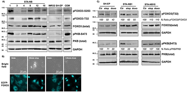
A panel of patient-derived NB cell lines (STA-NB1, STA-NB3, STA-NB4, STA-NB8, STA-NB12, STA-NB15) and the cell lines SH-EP and IMR32 were analyzed for the expression and phospho-status of FOXO3, PKB, pFOXO3-T32, pFOXO3-S253, and pPKB-S473 by immunoblot. CCRF-CEM leukemia cells were included as a positive control for PKB-hyperactivation A. SH-EP/ECFP-FOXO3 cells expressing ECFP-tagged FOXO3 were treated with etoposide (20 μg/ml) for 90 minutes or with doxorubicin (0.25 μg/ml) for 120 minutes and monitored by live cell fluorescence imaging B. SH-EP, STA-NB1 and STA-NB15 cells were treated with etoposide (10 μg/ml) and doxorubicin (0.5 μg/ml) for 2 and 6 hours, respectively, and subjected to immunoblot analyses using antibodies directed against FOXO3, pFOXO3-T32, PKB, pPKB-S473 and GAPDH as a housekeeping control C.