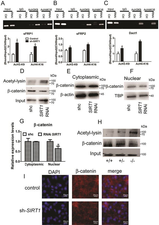
Figure 5. SIRT1 regulates Wnt signaling by deacetylating histones and β-catenin.

A, B and C. Pooled populations of C3H101/2 cells stably selected to express SIRT1 RNAi constructs were analyzed via ChIP. ChIP was performed with antibodies acetylated histone H3, lysine 9 (H3K9) and H4, lysine 16 (H4K16), or with IgG antibody controls. Promoter sequence was amplified by RT-PCR under linear conditions for the genes sFRP1 (A), sFRP2 (B) and Dact1 (C). The acetylation of H3K9 and acetylation of H4K16 at the sFRP1 (A), sFRP2 (B) and Dact1 (C) promoters were measured by Real-time PCR. Bars indicate SD. *P < 0.05. n = 3 per group. D. After stably transfected with SIRT1 RNAi plasmid in C3H101/2 cells. Endogenous β-catenin was immunopreciptated with anti-β-catenin, and acetylated β-catenin was identified using anti-acetyl-lysine. E. The protein levels of β-catenin in cytoplasms were measured by immunoblotting. F. The protein levels of β-catenin in nuclear were measured by immunoblotting. G. The relative expression protein levels of β-catenin in cytoplasmic and nuclear. Bars indicate SD. *P < 0.05. n = 3 per group. H. Pooled populations of of differentiated MEF cells were analyzed via IP. β-catenin was immunopreciptated with anti-β-catenin, and acetylated β-catenin was identified using anti-acetyl-lysine. I. Immunofluorescent staining of DAPI and β-catenin in control and SIRT1 RNAi C3H101/2 cells.
