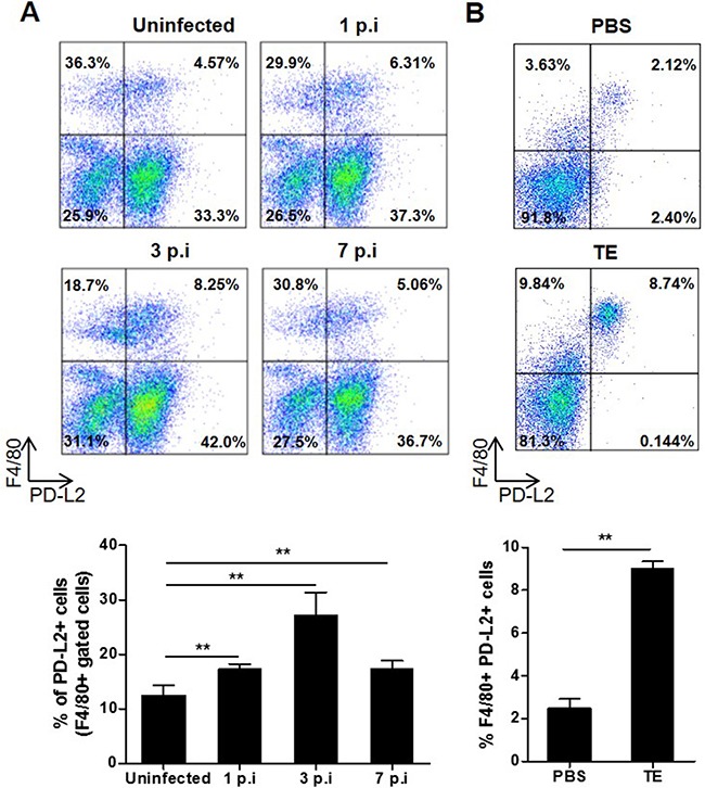
Figure 1. Effect of F. hepatica infection or total extract antigen injection on PD-L2 expression by peritoneal macrophages.

A. BALB/c mice were infected with 8 F. hepatica metacercariae, and PC were isolated at days 1, 3 and 7 p.i. Uninfected animals were analyzed in parallel as controls. PC were processed by flow cytometry and analyzed for F4/80+PD-L2+ cells. Representative dot plots of each group of mice are shown, with bars displaying the percentage of PD-L2+ cells gated on F4/80+ cells, ** p< 0.005. B. BALB/c WT mice were injected with 80 μg of TE or PBS as control and the PC were obtained 24 h later. F4/80+ PD-L2+ cells were analyzed by flow cytometry. A representative dot plot for each group of mice is shown. Bars display percentage of F4/80+ PD-L2+ cells, **p=0.0038. Data are representative of two independent experiments and expressed as mean ± SD.
